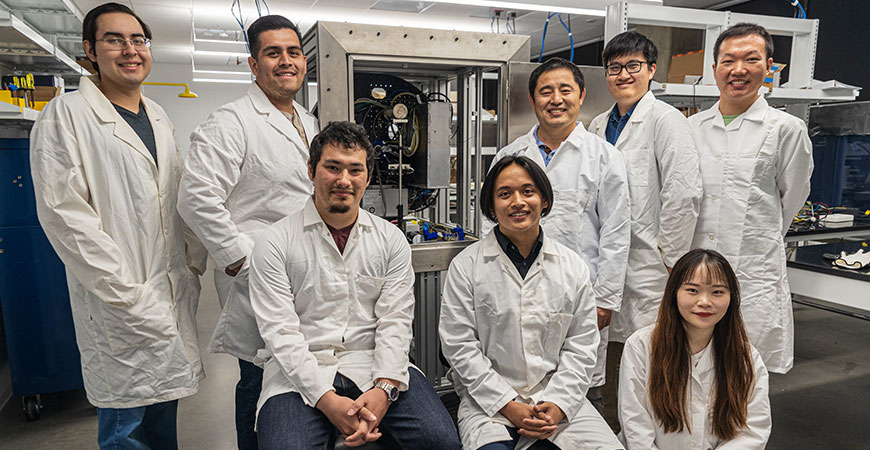
Bioengineering Professor Changqing Li is building a high-resolution CT imaging scanner that will allow scientists to study and understand how oxygen plays a role in cancer therapy and stem cells growing in deep tissue such as bone marrow, and possibly develop new advances to culture stem cells outside the body and therapeutics to control tumor growth.
Funded by grants from the National Institutes of Health (NIH), Li and fellow bioengineering Professor Joel Spencer are working with molecules that, when excited by the X-ray beam, emit light in the visible spectrum. By measuring the intensity of the light and how long it takes to be emitted, the researchers can detect how much oxygen is present in the tissue.
The project, called Bio-tissue Oxygenation Nanophosphor Enabled Sensing (BONES), would be a brand-new medical imaging technique with an unprecedented combination of chemical sensitivity and high-spatial resolution imaging through deep tissue.
The industry-academia partnership grant provides about $1.9 million under the Small Business Technology Transfer program. By partnering with Bay Area company Sigray Inc., the researchers use the power of a bright X-ray tube and a fancy X-ray optics focusing a superfine X-ray beam — to peer through thick tissue.
Hypoxic (low oxygen) conditions affect many medical conditions such as cancer, chronic kidney disease and failed organ transplant, but the heterogeneous nature of hypoxia is not well understood, Li said.
For example, bone marrow is a particularly hypoxic tissue, and its low-oxygen environment enables bone marrow to maintain adult stem cells. But the same conditions are believed to harbor cancer cells, which is why bone is a common cancer metastasis site.
Both Li and Spencer focus on biomedical imaging. Spencer had already developed a technique for visualizing stem cells in live, intact mice, but Li’s idea for BONES would allow researchers to see even deeper inside to directly measure molecular oxygen.
“It’s not easy to measure oxygenation in deep tissue,” Li said. “Right now, we can use a needle to extract samples, but it can only study that one spot. The proposed novel technology, BONES, will let us see inside a whole area of tissue such as tumors and bone marrow.”
Changes in the oxygenation levels of tumors can be indicative of responses to therapies, Spencer said. And in bone marrow, he explained, oxygenation is important for the tissue’s health.
“That can also be in the context of treatment, such as with bone marrow transplants,” he said. “You’d want to look at the recovery of the bone marrow. Oxygenation changes during recovery, but right now, no one has a way to look deep into the center of the marrow.”
Besides being colleagues in the Department of Bioengineering, both Li and Spencer are affiliated with the Health Sciences Research Institute. The work on BONES under this grant lasts through 2024. It builds on work Li has been doing since he joined the campus in 2012, including through a $2.5 million R01 grant from the NIH to develop a first-of-its kind X-ray luminescence tomography scanner that allows researchers to visualize how cancer progresses and monitor the effectiveness of novel drug-delivery systems in live animals — without invasive surgeries or euthanizing the animals.

